


Podiatry Ultrasound Guide for Foot and Ankle Pain
A painful first step in the morning, swelling around the ankle after a run, or a forefoot pain that keeps returning can each have several possible causes. A podiatry ultrasound guide is useful because it explains how a scan can add clarity to a clinical assessment, rather than leaving treatment to guesswork. For many foot and ankle problems, the most valuable part is being able to examine the painful structure, see it in real time and discuss the findings during the same appointment.
What podiatry ultrasound is used for
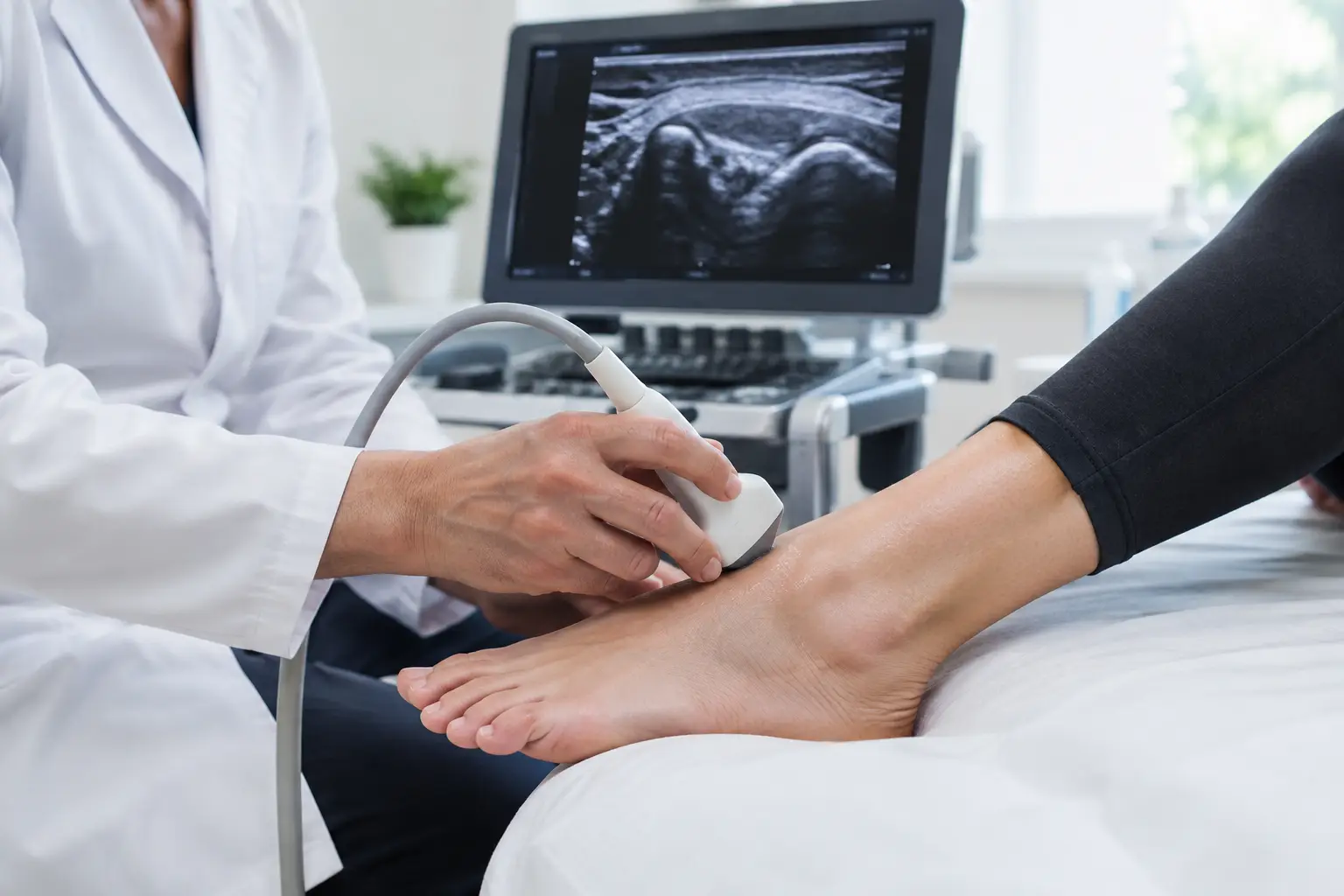
Diagnostic ultrasound uses sound waves to create moving images of soft tissues beneath the skin. It does not use radiation and is usually comfortable, quick and non-invasive. A small amount of gel is applied to the area, then a handheld probe is moved over the foot, ankle or lower leg to assess the relevant structures.
In podiatry, ultrasound is particularly helpful for examining tendons, ligaments, plantar fascia, bursae, joints, nerves and areas of fluid or soft-tissue swelling. It can show whether a tendon is thickened, whether the plantar fascia has changes consistent with plantar heel pain, or whether inflammation is present around a joint or tendon sheath.
The scan is not intended to replace a proper assessment. Symptoms, medical history, footwear, activity levels, walking pattern and physical examination all matter. A finding on a scan only becomes clinically meaningful when it matches the location of pain and the way symptoms behave. Some changes can be seen in people without pain, particularly in tendons, so treatment should never be based on an image alone.
When an ultrasound scan may help
Ultrasound is often considered when pain is persistent, recurring or not responding as expected to initial care. It may also be appropriate when the diagnosis is uncertain, when a specific soft-tissue injury is suspected, or when the result would alter the treatment plan.
Common reasons for assessment include heel pain that has not settled, pain around the Achilles tendon, swelling on the top or side of the foot, suspected Morton’s neuroma, pain beneath the ball of the foot, ankle tendon pain and symptoms following a twist or sporting injury. It may also be used to assess a lump, a ganglion or inflammation around a joint.
For an active person, the question is often whether it is safe to continue running or training. For someone whose work involves long periods on their feet, the priority may be reducing pain without unnecessary time away from normal activity. Ultrasound can help identify the tissues involved, but advice still depends on the severity of symptoms, function and the demands placed on the foot.
Conditions that ultrasound can assess well
Ultrasound is particularly effective for superficial soft tissues. In the right clinical context, it can support assessment of plantar fasciopathy, Achilles tendinopathy, peroneal or tibialis posterior tendon problems, bursitis, synovitis, Morton’s neuroma and some ligament injuries. It can also identify fluid collections, tendon sheath inflammation and certain forms of soft-tissue thickening.
Its real-time nature is a practical advantage. The clinician can scan the exact point that reproduces symptoms, compare the affected side with the other foot where useful, and observe a tendon or joint while it moves. This dynamic assessment can provide information that a static image cannot.
What ultrasound cannot show completely
No scan answers every question. Ultrasound has limitations when assessing structures deep inside the foot, and it is not the best test for every suspected injury. If a stress fracture, bone injury, significant joint damage or a problem involving deeper tissues is suspected, an X-ray, MRI or other investigation may be more appropriate.
Ultrasound also cannot determine the full cause of every pain presentation. Nerve-related symptoms can arise from the back, leg or local foot structures. Pain beneath the forefoot may be influenced by joint loading, footwear and foot mechanics as well as local inflammation. A careful assessment prevents a scan result from being overinterpreted.
What happens during a podiatry ultrasound appointment
A well-structured appointment begins with questions before the probe is used. Your podiatrist will ask when the symptoms started, what makes them worse or better, whether there was an injury, and how the issue affects walking, work, exercise and footwear choices. Relevant health conditions and previous treatments are also considered.
This is followed by examination. The foot and lower limb may be assessed while standing, walking and sitting. Your clinician will check the painful area, joint movement, muscle and tendon function, swelling and any signs that suggest a different diagnosis. If ultrasound is clinically indicated, the scan then becomes part of the same assessment rather than a separate service disconnected from your symptoms.
During the scan, you may be asked to move your ankle, toes or foot. You may also be asked to point to the precise site of discomfort. Most scans take only a short time, although the overall appointment allows time for assessment, explanation and treatment planning.
The findings should be discussed in plain language. That includes what can be seen, what it is likely to mean, and what remains uncertain. A useful consultation does not simply label a structure as abnormal. It explains whether the finding is likely to be responsible for your pain and what can realistically help it settle.
How scan findings guide treatment
The purpose of imaging is to make treatment more precise. If the assessment supports plantar fasciopathy, for example, management may include load modification, footwear advice, stretching or strengthening, manual therapy, taping, orthoses or shockwave treatment where appropriate. The right combination depends on the duration of symptoms, tissue irritability and your activity goals.
For tendon problems, treatment often focuses on progressive rehabilitation. This may involve temporary changes to running or walking volume, a structured strengthening programme and practical support to reduce excessive strain while capacity is rebuilt. Complete rest is not always the answer, particularly for persistent tendon pain. Equally, pushing through sharp or worsening pain can delay recovery.
Where a local inflammatory condition is confirmed and simpler measures have not been effective, an injection may be discussed. Ultrasound can be used to guide certain injections accurately to the intended area. However, an injection is not automatically the best next step. The potential benefit, alternatives, risks and need for rehabilitation should be considered carefully before proceeding.
Orthotic devices may be recommended when foot mechanics or loading patterns are contributing to symptoms. They are not a universal solution, and they work best when prescribed for a clear reason as part of a wider plan. The scan may identify the affected tissue, while the physical assessment helps establish why it is being overloaded.
Getting the most from your assessment
Bring or wear the footwear you use most often, especially running shoes, work shoes or boots that may be relevant to your symptoms. If you exercise, it is helpful to describe recent changes in distance, pace, terrain, gym work or sport. Small changes in training load can be significant when symptoms develop.
Be clear about the pattern of pain. Is it worst with the first few steps, during activity, after activity or at night? Does it feel sharp, burning, stiff or unstable? These details guide the assessment and can be as useful as the scan itself.
If you have had previous imaging, reports or treatment, share these where possible. A prior diagnosis should be reviewed rather than assumed to remain correct, particularly if symptoms have changed or failed to improve.
When prompt medical assessment matters
Most foot and ankle pain can be assessed through a planned appointment, but some symptoms need urgent attention. Seek prompt medical advice for a suddenly hot, red and swollen joint, a suspected infection, a foot that is numb or cold, severe pain after significant trauma, inability to bear weight, or an open wound. People with diabetes, poor circulation or reduced sensation should also seek early assessment for new wounds, colour changes or unexplained swelling.
For persistent but non-urgent pain, a consultation-led approach offers a sensible next step. At South London Foot Clinic, ultrasound is used when it will genuinely improve diagnostic clarity and help direct treatment. The aim is not simply to produce an image, but to give you a clear explanation of the problem and a practical route back to comfortable movement.


