


Is Ankle Ultrasound Worth It for Lasting Pain?
A sore ankle that has not settled after a twist, a run or a gradual increase in activity can be difficult to interpret. The swelling may have reduced, yet pain remains when walking downstairs, pushing off, turning, or wearing certain shoes. If you are asking, is ankle ultrasound worth it, the useful question is whether a scan is likely to change the diagnosis or treatment plan.
For many people with ongoing ankle pain, it can. Ultrasound provides a real-time view of tendons, ligaments and other soft tissues around the ankle. Used alongside a structured clinical examination, it can help identify the source of symptoms and avoid treatment based on assumption alone. It is not required for every ankle problem, and it is not a substitute for a thorough assessment. Its value comes from using it at the right point, for the right clinical question.
When is ankle ultrasound worth it?
An ankle ultrasound is often worthwhile when symptoms are persistent, recurrent or unclear after an initial injury. This includes pain that has continued beyond the expected early recovery period, repeated ankle sprains, localised swelling, clicking, weakness or pain that returns as soon as activity resumes.
It can be particularly helpful when examination suggests a tendon problem. The tendons around the ankle work hard to stabilise the foot and transfer load during walking, running and changes of direction. Irritation, thickening, small tears or altered movement of a tendon can cause very specific symptoms, but these problems are not always distinguishable from a simple sprain without imaging.
A scan may also be clinically indicated if there is concern about the Achilles tendon, peroneal tendons on the outside of the ankle, tibialis posterior tendon on the inside of the ankle, or tendons across the front of the joint. It can assess fluid around a tendon, changes in tendon structure and, in some cases, whether a tendon moves abnormally during movement.
The benefit is not simply having a picture of the ankle. The benefit is connecting what appears on the scan with where you feel pain, what activities provoke it, how the joint moves and how you walk. This is why ultrasound is most useful as part of a consultation-led assessment rather than as a standalone test.
What an ankle ultrasound can show
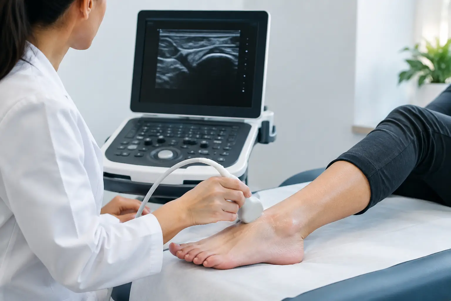
Ultrasound uses sound waves to create images of soft tissue. There is no radiation, and the examination is usually comfortable. A small amount of gel is applied to the skin, then the clinician moves a probe over the area being assessed.
Unlike a static scan, ultrasound can be performed dynamically. The clinician can ask you to move your foot, point your toes, turn the sole in or out, or reproduce a movement that causes discomfort. This can be valuable for tendon instability, impingement symptoms and pain that only occurs under movement or load.
Depending on the clinical question, an ankle ultrasound can help assess:
- tendon thickening, degeneration, inflammation or partial tears
- fluid within a tendon sheath or around the joint
- ligament injury in selected areas
- bursae, ganglion cysts and other soft-tissue lumps
- some signs of joint inflammation or synovitis
- areas of increased blood flow that may support evidence of active tissue irritation
It may also help guide a procedure where this is clinically appropriate. For example, if an injection is being considered after examination and discussion, ultrasound guidance can improve precision by confirming the relevant tissue and target area.
What it cannot tell you
A clear answer is just as important as recognising the limitations. Ultrasound does not show every structure in the ankle equally well. Bones are not assessed in the same way as on an X-ray, so it is not the first investigation when a fracture, stress fracture or bony alignment problem is suspected. It may also be less suitable than MRI for certain deep joint structures, cartilage injuries, bone marrow changes or complex ligament damage.
This does not mean MRI is automatically the better choice. MRI provides detailed information but is not always the most practical or necessary first investigation. If the suspected issue is superficial and dynamic, such as a tendon disorder, ultrasound may answer the question more directly and can be performed during the assessment.
A scan must also be interpreted carefully. Structural changes can sometimes be present without causing pain, particularly in active adults or people with a history of previous injury. Conversely, an ankle can be painful despite limited visible changes on imaging. Good clinical care considers the scan findings in context rather than treating an image.
What happens during an ultrasound-supported ankle assessment?
The appointment should begin with your history. Your clinician will ask how the problem started, where symptoms are felt, whether swelling or bruising occurred, which movements are limited and what has or has not helped. Your work, footwear, sport, training load and medical history can all be relevant.
A physical examination then assesses tenderness, range of motion, joint stability, strength and the way the foot and ankle behave under load. In some cases, this examination gives a sufficiently clear diagnosis and imaging is not needed. In others, ultrasound is used to confirm or refine the clinical impression.
At South London Foot Clinic, this approach allows consultation, imaging where indicated and discussion of the findings to take place within a structured diagnostic pathway. You should leave with an explanation of what has been found, what it is likely to mean for recovery and what the sensible next step is.
That plan may involve activity modification, targeted rehabilitation, footwear advice, taping or bracing, custom orthotics, manual treatment or a procedure such as an injection where appropriate. The treatment should follow the diagnosis, not precede it.
When a scan may not be necessary
Not every ankle injury needs ultrasound. A mild sprain that is steadily improving, with normal function returning and no concerning symptoms, can often be managed conservatively without imaging. Similarly, a scan is unlikely to add much where the clinical examination already provides a clear explanation and would not alter treatment.
Urgent assessment is more appropriate than routine ultrasound if you cannot bear weight after an injury, have obvious deformity, severe swelling, numbness, a cold or discoloured foot, fever with a hot swollen joint, or calf swelling and breathlessness. These symptoms can require prompt medical investigation.
If pain is persistent but the scan is normal, that is still useful information. It may shift attention towards joint mechanics, load management, referred pain, nerve irritation or a condition better assessed through a different type of imaging. A normal result does not mean your symptoms are dismissed. It helps narrow the possibilities and guide the next decision.
Is the cost of an ankle ultrasound justified?
The cost is easier to justify when it prevents weeks or months of trial-and-error treatment. Repeatedly resting an ankle, changing trainers or trying exercises without knowing the affected structure can be frustrating, particularly if symptoms keep returning. For runners, people who stand for work and those preparing for a sporting event or holiday, diagnostic clarity can be especially valuable.
However, the scan is not worthwhile merely because it is available. The right test is one that answers a specific question and has a realistic chance of changing management. Before proceeding, it is reasonable to ask what the clinician is looking for, whether the result will alter the plan and whether another investigation may be more suitable.
For many persistent ankle problems, ultrasound offers a practical balance of detail, speed and clinical relevance. It can show how tissues behave while you move, provide immediate discussion of the findings and support a more targeted recovery plan. If your ankle pain has stopped improving or keeps returning, a properly assessed scan can be the step that replaces uncertainty with a clear, realistic route back to comfortable movement.


