


Forefoot Pain Orthotics Case Study in Practice
Pain beneath the ball of the foot is often described as though it has one obvious cause: “metatarsalgia”. In practice, that word only describes the location of symptoms. A proper forefoot pain orthotics case study shows why identifying the structure under load matters before deciding whether an orthotic is likely to help.
The following is an illustrative composite case based on a common clinical presentation. It is not a substitute for an individual assessment, but it demonstrates the structured process used to move from persistent forefoot pain to a diagnosis-led treatment plan.
The presenting problem
A 46-year-old recreational runner attended with pain under the second and third metatarsal heads of the right foot. The discomfort had started gradually over several months and was initially only noticeable after longer runs. It had progressed to pain during ordinary walking, particularly in thin-soled shoes and when barefoot on hard floors.
The patient described a feeling of pressure and occasional burning beneath the forefoot rather than a sharp pain between the toes. There was no single injury, no visible swelling and no previous history of fracture. They had tried changing trainers, reducing running mileage and using an off-the-shelf gel forefoot pad. These measures offered short-lived relief but did not address the problem.
This history immediately raises several possibilities. Pain in this area can relate to overload of the metatarsal heads, inflammation around a joint, plantar plate injury, stress reaction, bursitis, nerve irritation or a combination of factors. A generic insole may feel comfortable, but comfort alone does not establish whether it is applying pressure in the right place.
Assessment before prescribing orthotics
The first stage was a detailed consultation and lower-limb examination. The aim was not simply to locate where the patient felt pain, but to understand why that part of the foot was being overloaded.
Examination identified local tenderness beneath the second metatarsophalangeal joint, with discomfort on loading the joint and mild reduction in ankle dorsiflexion. The second toe showed early elevation compared with the adjacent toes, suggesting reduced control around the joint. There was no marked numbness on examination and no pain reproduced by compression of the forefoot, making an interdigital neuroma less likely at that stage.
Standing assessment showed a relatively mobile foot with increased pressure passing through the central forefoot during late stance. Gait observation suggested that limited ankle movement was encouraging an earlier heel lift, transferring load forwards sooner and for longer. This did not mean that the ankle was the sole cause of the symptoms. It did, however, help explain why the second metatarsal area was repeatedly being asked to absorb more force.
Where clinically indicated, diagnostic ultrasound can add useful clarity. In this case, ultrasound was used within the consultation to assess the painful area directly. It demonstrated irritation around the second metatarsophalangeal joint and changes consistent with plantar plate stress, without evidence of a complete tear. The findings supported a mechanical overload diagnosis and helped rule out some alternative treatment pathways.
Why the diagnosis changed the treatment plan
It would have been easy to label the symptoms as “metatarsalgia” and provide a standard forefoot cushion. That approach may reduce impact temporarily, but it may also fail to alter the loading pattern that is maintaining joint irritation.
The clinical working diagnosis was second metatarsophalangeal joint synovitis with early plantar plate overload. The treatment priorities were therefore to reduce pressure beneath the affected area, improve support to the forefoot, limit excessive strain through the joint and address the restricted ankle movement contributing to early forefoot loading.
This distinction is particularly important with orthotics. An orthotic is not simply an item placed inside a shoe. Its effectiveness depends on the diagnosis, foot shape, shoe type, activity demands and the way the device is adjusted over time. For some causes of forefoot pain, an orthotic may be central to management. For others, it may be only one part of treatment or may not be appropriate at all.
Forefoot pain orthotics case study: the device design
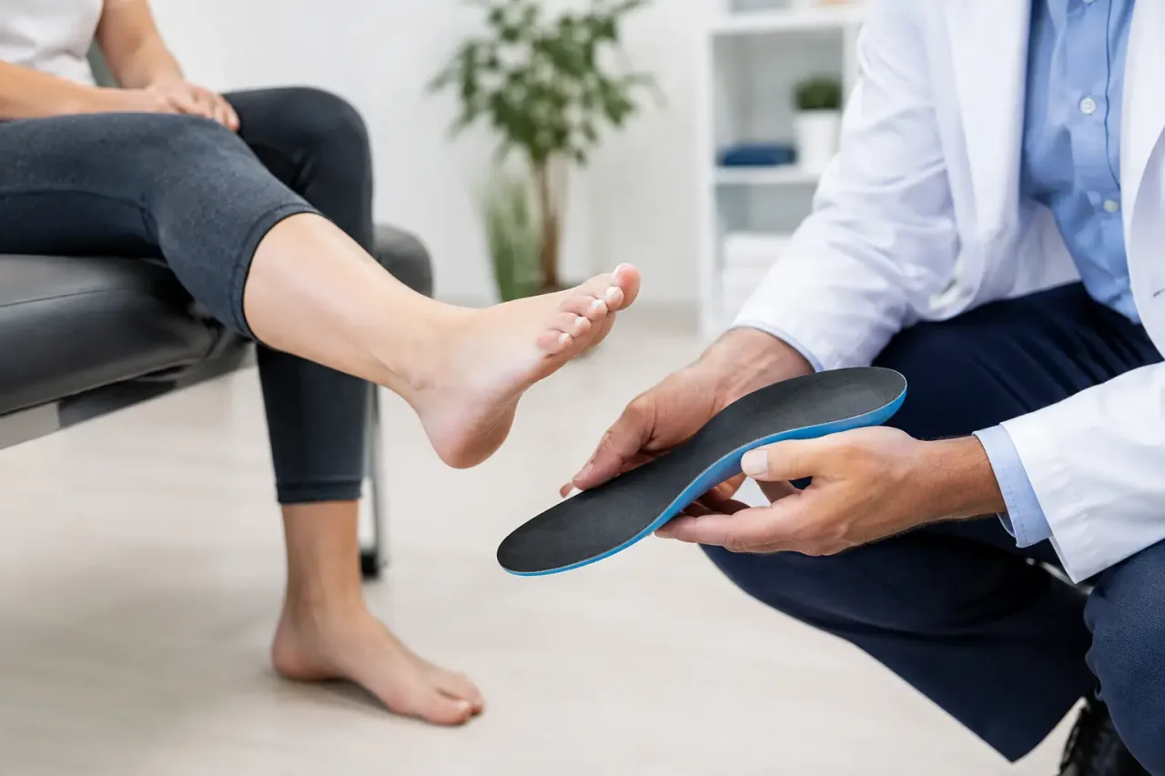
A custom orthotic was prescribed following assessment rather than selected from a standard template. The device was designed to provide stable rearfoot and midfoot support while accommodating the forefoot. The key feature was a carefully positioned metatarsal dome placed just behind the painful metatarsal heads.
The purpose of the dome was not to press directly into the sore area. Positioned correctly, it supports the metatarsal shafts and can redistribute load away from the painful joint region during propulsion. It is a small adjustment with a significant practical consequence: if it is too far forward, it can increase discomfort; too far back, and it may have little effect.
A local accommodation was also incorporated beneath the sensitive second metatarsal head. The patient was advised that the orthotics would need to be worn in suitable footwear with adequate depth and a removable insole where possible. Very narrow, shallow or flexible shoes can prevent an orthotic from sitting correctly and may limit its benefit.
The device was introduced gradually over the first one to two weeks. This matters because the patient was not only adapting to new support under the foot, but also changing a movement pattern that had developed over months. Immediate full-time use can be uncomfortable for some people, particularly where tissues are already sensitive.
Rehabilitation and load management
Orthotics reduced the mechanical stress, but they were not presented as a standalone cure. The patient was given a practical rehabilitation plan focused on calf flexibility, ankle dorsiflexion and controlled strengthening of the intrinsic foot muscles. Exercises were selected to support the treatment goal rather than added as a generic list.
Running was modified rather than stopped indefinitely. For the first phase, longer runs and speed sessions were reduced, while cycling and strength work were used to maintain fitness. The patient was advised to monitor symptoms during activity and the following morning. Mild discomfort that settled promptly could be acceptable; worsening pain, altered gait or increasing next-day symptoms indicated that the load was too high.
Footwear was also reviewed. The patient did not need an unusually rigid shoe, but benefited from a trainer with adequate forefoot width, cushioning and enough internal volume for the orthotic. A slightly rocker-shaped sole may help some patients by reducing demand through the painful forefoot during toe-off. It is not necessary or suitable for everyone, which is why shoe advice should follow examination rather than fashion or marketing claims.
Review and outcome
At a six-week review, the patient reported substantially less pain during walking and could tolerate short, easy runs without the previous sharp increase in symptoms. There was still mild tenderness after prolonged activity, but the baseline pain had reduced and the patient no longer needed to avoid hard flooring at home.
The orthotics were reviewed in the shoes most commonly worn. A minor adjustment was made to refine the accommodation beneath the second metatarsal area. This is a normal part of custom orthotic care. The initial device is based on clinical findings, but response to treatment provides further information about how the foot behaves under real-life conditions.
By 12 weeks, the patient had returned to a progressive running programme and was managing day-to-day activity comfortably. The outcome was not attributed to one component alone. It reflected a combination of accurate diagnosis, pressure redistribution, footwear changes, rehabilitation and realistic control of training load.
When orthotics may not be the first answer
Orthotics are often helpful for mechanical forefoot pain, but they should not be prescribed automatically. Sudden severe pain, marked swelling, bruising, night pain, loss of sensation, signs of infection or an inability to bear weight need prompt clinical assessment. A stress fracture, significant plantar plate tear, inflammatory joint condition or nerve problem may require a different pathway.
Even when an orthotic is clinically appropriate, results depend on the cause of pain and how consistently the treatment plan can be followed. A device cannot fully offset shoes that compress the forefoot, a rapid increase in running volume or an untreated joint injury. Equally, persistent symptoms do not always mean that orthotics have failed. They may signal that the diagnosis needs to be revisited, the device needs adjustment or another treatment is required.
At South London Foot Clinic, the focus is on establishing what is causing the pain before recommending treatment. When forefoot symptoms are assessed carefully, with ultrasound used where it can answer a clinical question, orthotics can become a precise part of a wider recovery plan rather than a trial-and-error purchase.
Persistent pain beneath the ball of the foot deserves more than a generic pad. A clear diagnosis gives you a better basis for choosing treatment, returning to activity and knowing what to do if symptoms do not settle as expected.


