


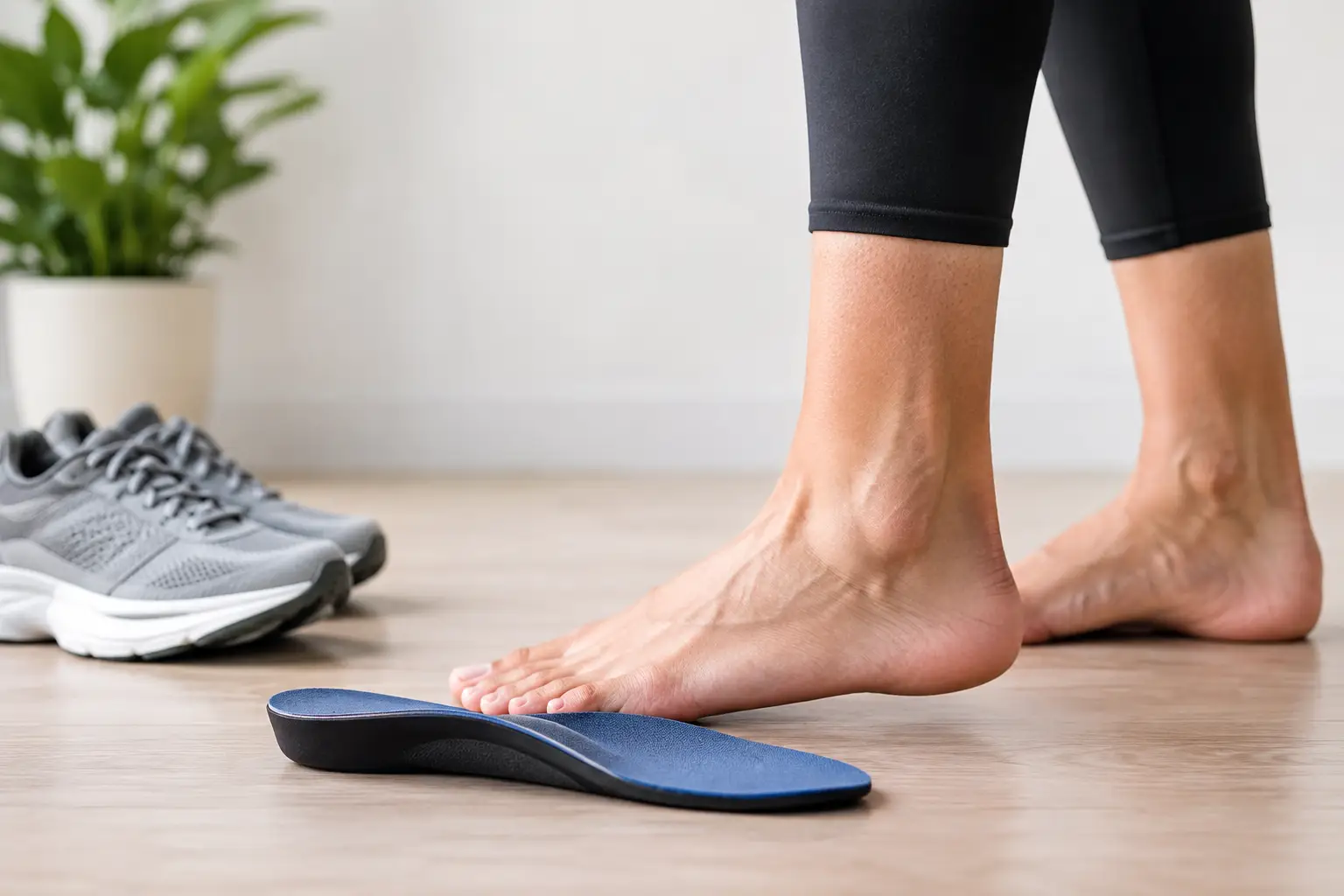
Adult Flat Feet Guide for Pain and Support
Flat feet are not automatically a problem. Many adults have low arches, have always had them and remain completely comfortable. The concern begins when a change in foot shape is accompanied by pain, fatigue, reduced walking tolerance or recurring ankle and knee symptoms. This adult flat feet guide explains when flat feet need assessment, what may be causing them and how treatment is planned around your individual diagnosis.
What flat feet can mean in adults
A flat foot describes a low or absent arch when standing. In some people, the arch reappears when they sit, rise onto their toes or take weight off the foot. This is often called a flexible flat foot. In others, the foot remains flat and may become stiff, which can indicate a more established structural change.
The visible shape of the foot is only one part of the picture. Some people have a low arch without symptoms, while another person with a similar-looking foot may experience pain along the inside of the ankle, under the arch, in the heel or through the forefoot. The way the foot moves, the condition of the tendons and joints, footwear, activity levels and previous injuries all matter.
Adult-acquired flatfoot is different from simply having naturally low arches. It can develop over time and may become more pronounced on one side. In clinical terms, this is often associated with posterior tibial tendon dysfunction or progressive collapsing foot deformity. The posterior tibial tendon supports the arch and helps control the foot during walking. If it becomes irritated, weakened or torn, the arch may gradually lower and the heel may drift outwards.
Adult flat feet guide: symptoms that merit attention
Pain is the key reason to seek advice, particularly if it is new, persistent or limiting normal activity. Common patterns include pain or swelling behind and below the inside ankle bone, aching through the arch, discomfort in the outer ankle as the foot changes position, or tired feet after standing and walking.
You may also notice that one shoe wears differently, you need wider footwear than before, or you can no longer comfortably perform a single-leg heel raise on the affected side. Runners may report a gradual loss of control or endurance rather than one clear injury. For others, a long day on their feet becomes increasingly difficult.
A prompt assessment is sensible if any of the following apply:
- The arch on one foot has noticeably lowered or changed shape.
- Ankle or arch pain has not settled with a short period of reduced activity and supportive footwear.
- Swelling, warmth or tenderness is present around the inside ankle.
- You have diabetes, reduced sensation, inflammatory arthritis or a history of significant foot or ankle injury.
- The foot has become stiff, painful at rest or difficult to fit into usual shoes.
Sudden inability to bear weight, marked redness or heat, a wound, fever, or a painful swollen foot with reduced sensation requires urgent medical advice. People with diabetes should not wait for a routine appointment if there is a new break in the skin, colour change or rapidly developing swelling.
Why arches can lower later in life
There is rarely one single cause. Repeated load can irritate the posterior tibial tendon, particularly in people who walk or stand for work, have recently increased running or exercise, or are returning to activity after a period of inactivity. A previous ankle sprain can alter how the foot and ankle are controlled, even when the original injury seemed to recover.
Age-related tendon change, joint wear and inflammatory conditions can also play a part. Body weight is relevant because it increases the load through the foot, but it is not a complete explanation and should not be treated as one. Foot shape, calf tightness, footwear, strength and the demands of daily life can all contribute.
Pregnancy can temporarily affect foot shape and ligament laxity. Some people also notice more symptoms after a change in job, a house move or a holiday involving much more walking than usual. The useful question is not simply, “Do I have flat feet?” It is, “Why is this foot painful now, and which structures are under strain?”
A structured assessment gives clearer answers
A useful appointment starts with the history. Your clinician will ask where the pain is felt, when it began, what makes it worse, whether there has been a change in activity or footwear, and how the issue affects work, exercise and everyday walking.
The examination then considers more than the arch. It may include observation of standing alignment and gait, joint movement, calf flexibility, strength testing, tendon tenderness and the ability to rise onto the toes. Both feet are assessed because differences between sides are often clinically meaningful.
Diagnostic ultrasound may be appropriate when symptoms suggest a tendon, ligament, bursa or other soft-tissue problem. It can help assess the posterior tibial tendon for thickening, inflammation or tearing, and it allows the clinician to relate imaging findings to the exact site of symptoms. Ultrasound does not replace a careful examination, and it is not needed in every case. Its value is in answering a specific clinical question and helping to guide treatment.
X-rays, MRI or onward referral may be recommended where there is concern about arthritis, a stress injury, a rigid deformity, a more significant tendon injury or another condition that ultrasound cannot fully assess. Clear diagnosis prevents the common mistake of treating every painful flat foot with the same insole.
Treatment depends on the cause and stage
Early treatment often aims to reduce stress on irritated tissues while restoring the foot’s ability to tolerate load. This may involve a temporary change to activity, better footwear, targeted rehabilitation and appropriate support. The aim is not necessarily to create a high arch. It is to improve comfort, control and function.
Footwear should have a stable heel counter, enough depth and width for the foot, and a sole that does not twist excessively through the middle. Very soft, worn or unsupportive shoes can make symptoms harder to settle. However, a stiff shoe is not right for everyone, particularly where forefoot arthritis or restricted ankle movement is present.
Orthoses can be helpful when they are prescribed for a defined reason. They may support the arch, influence heel position, redistribute pressure or reduce strain on a painful tendon. Off-the-shelf devices are sufficient for some straightforward cases. Custom orthotics may be considered where foot shape, symptoms, gait findings or previous treatment response indicate that more specific control is needed. They work best alongside rehabilitation and appropriate footwear, rather than as a stand-alone solution.
Rehabilitation commonly focuses on calf flexibility, balance, foot control and progressive strengthening of the calf and supporting muscles around the ankle. Exercises must be matched to the diagnosis. If a tendon is significantly irritated, aggressive strengthening too early can prolong symptoms. Conversely, prolonged rest without rebuilding capacity can leave the foot vulnerable when normal activity resumes.
Where pain is persistent and imaging confirms inflammation in a suitable structure, injection treatment may occasionally be discussed. This is a decision made after considering the tissue involved, the likely benefit, risks and the wider rehabilitation plan. Steroid injections are not appropriate for every tendon problem, and symptom relief alone does not resolve the underlying loading issue.
More advanced flatfoot deformity, marked stiffness or significant tendon damage may need assessment by an orthopaedic foot and ankle specialist. Surgery is not the starting point for most people, but timely referral matters when the foot is progressively changing shape or conservative care has not provided adequate function.
What you can do while awaiting assessment
Choose the most supportive comfortable footwear you already own and avoid repeatedly testing pain through long walks, runs or high-impact exercise. Reducing aggravating activity for a short period is sensible, but complete inactivity is rarely the long-term answer. Low-impact options, such as cycling or swimming, may be more comfortable while the cause is being established.
Keep a brief note of where pain occurs, how long it lasts after activity and whether there is swelling. Bring the shoes you wear most often to an appointment if they show unusual wear. These details can help identify a pattern that is not obvious during a short consultation.
Persistent pain should not be dismissed as a normal consequence of ageing or simply “fallen arches”. At South London Foot Clinic, assessment can combine examination with diagnostic ultrasound when clinically indicated, followed by a clear discussion of the findings and realistic treatment options. The right next step is the one that matches the structure causing your symptoms, your day-to-day demands and the level of support your foot genuinely needs.


